On Friday, October 26th, I made my first observation of the MicroAquarium since we put them together in class. The first thing I noticed was that the water level had visibly gone down. At first glance it appeared as if the previously unidentified bug and plants were still alive.
.JPG) |
| Figure 1. The water level had dropped quite a bit since the MicroAquarium was constructed. |
Luckily the lab was not very busy and I immediately had access to a microscope. The microscope at my particular station was a Leitz Laborlux 11 with an attached camcorder and remote for taking still images through the microscope. What seemed to be a pretty quiet and still aquarium was suddenly very busy looking under magnification. Some organisms were darting around quickly and getting a picture of them would prove to be particulaly tough. I noticed many
Vorticella (figure 2) throughout the water (Patterson 1996). Some were alone and some were in small groups. They were easy to find because they are attached to the plants with a recoil-able stalk.
 |
| Figure 2. Vorticella specimens were common throughout the MicroAquarium. |
As I continued observing the aquarium I was also able to see many examples of Rotifer Euchlanis (figure 3, figure 4) swimming around (Patterson 1996). They were never too far from plant life and were observed floating detritus in the aquarium water. They are fairly quick swimmers but would only do so in short bursts.
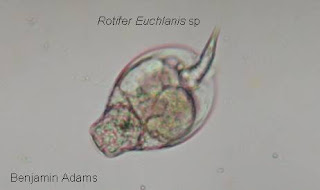 |
| Figure 3. A stationary Rotifer Euchlanis sp |
 |
| Figure 4. A Rotifer Euchlanis sp swimming. |
While scanning the water I noticed something that looked like a ball or a sphere. On further investigation I could see that it was covered in spines. It was motionless so I took advantage to take a good picture (figure 5). The organism was identified as
Acanthocystis sp (Patterson 1996). They are planktonic unicellular organisms and were found to be common in the water. I also happened to find a water flea (
Cyclidium sp) nearby on a plant (figure 6). I had seen many of them as I scanned the aquarium but they were usually moving much to fast to get a good picture.
 |
| Figure 5. Acanthocystis sp with visible spines. |
 |
| Figure 6. Cladocera sp, also known as a Water Flea. |
Many diatoms were also visible floating in the water. There were also many ciliates and flagellates swimming through the water. They were much too small and fast for me to coordinate a good picture at this particular station. This observation went very well although I wish I could have gotten some more pictures. I was happy to see my MicroAquarium was very lively and all seemed well in it's little environment.
.JPG)


.JPG)